Skull Theory Accuracy: Can Baby's Skull Shape Predict Gender?
Skull Theory Accuracy: Can Baby's Skull Shape Predict Gender?
Skull theory is one of the three most popular ultrasound-based gender prediction methods, alongside Ramzi theory and nub theory. It claims that the shape and features of a baby's skull on ultrasound can indicate whether you're having a boy or a girl.
But how accurate is skull theory really? Is there any scientific basis for it? And when should you use it versus other methods? Here's a thorough examination.
What Is Skull Theory?
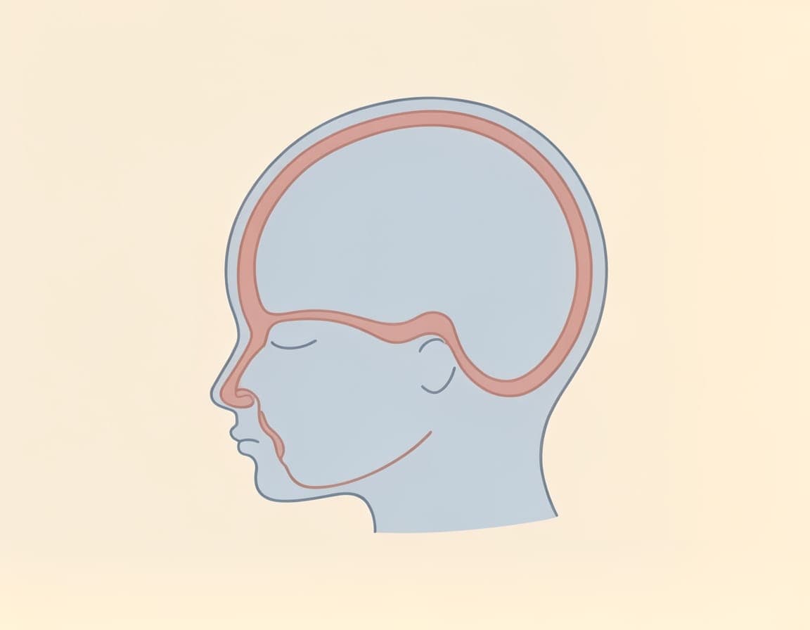
Skull theory analyzes the shape, contours, and features of a baby's cranium on ultrasound images to predict gender. The theory is based on observed differences between male and female skulls — differences that are well-documented in adult anthropology and forensic science.
The Key Indicators
According to skull theory, these features differ between boys and girls:
Male skull indicators:
- Blockier, more squared jaw and chin
- Prominent brow ridge (supraorbital ridge)
- Wider, more angular forehead
- Larger cranial volume relative to body size
- More defined occipital area (back of skull)
Female skull indicators:
- Rounder, more tapered jawline
- Smoother, less pronounced brow
- Rounder, higher forehead
- Smaller cranial volume relative to body size
- Softer occipital contour
The theory applies these adult anthropological differences to fetal ultrasound images, assuming that sexual dimorphism in cranial features begins developing before birth.
Skull Theory Accuracy: What the Evidence Shows
The Honest Assessment
Skull theory has the least scientific backing of the three major prediction methods. Here's what the evidence says:
- No peer-reviewed studies have specifically validated skull theory for fetal gender prediction
- The original anthropological data comes from adult skulls, not fetuses
- Fetal skull differences (if they exist) are likely subtle and difficult to detect on ultrasound
- Community surveys suggest accuracy closer to 50-70%, meaning it performs only slightly better than chance in many cases
Why the Evidence Is Limited
Several factors make skull theory difficult to validate:
-
Fetal skull development is ongoing. The skull bones are not fully formed or fused during pregnancy. The fontanelles (soft spots) are open, and the cranial bones are still developing their final shape.
-
Ultrasound image quality varies significantly. The subtle differences skull theory looks for can be easily obscured by poor image quality, baby's position, or the ultrasound machine's resolution.
-
Subjectivity of interpretation. Unlike nub theory, which measures a specific angle, skull theory relies on subjective visual assessment. Two different analysts may interpret the same image differently.
-
Individual variation is significant. Even among adult skulls, there is enormous overlap between male and female cranial features. Many female skulls have "masculine" features and vice versa.
Estimated Accuracy by Gestational Age
| Week | Estimated Accuracy | Reason |
|---|---|---|
| 12 weeks | 55-65% | Minimal cranial differentiation |
| 14 weeks | 60-70% | Slight ossification beginning |
| 16 weeks | 65-72% | More defined features |
| 20+ weeks | 70-78% | Better ossification, clearer images |
These estimates are based on practitioner experience and community data, not controlled studies.
How Skull Theory Compares to Other Methods
| Method | Best Timing | Estimated Accuracy | Scientific Basis |
|---|---|---|---|
| Nub Theory | 12-14 weeks | 80-92% | Some anatomical basis |
| Ramzi Theory | 6-12 weeks | 55-65% | Limited evidence |
| Skull Theory | 14+ weeks | 60-75% | Minimal evidence |
| NIPT Blood Test | 10+ weeks | 95-99% | Strong evidence |
| Anatomy Scan | 18-20 weeks | 95-99% | Medical standard |
Skull theory ranks below nub theory in accuracy but can still contribute useful information when combined with other methods.
When Skull Theory Works Best
Despite its limitations, skull theory can be valuable in specific situations:
1. As a Complementary Method
Skull theory shines when used alongside nub theory and Ramzi theory. If nub theory suggests a girl and skull theory also suggests a girl, the combined prediction carries more weight than either method alone.
2. When Other Methods Are Unavailable
Some ultrasound images don't show the genital tubercle clearly enough for nub theory, or the placenta position may be ambiguous for Ramzi theory. In these cases, skull features might still be visible and provide some predictive value.
3. Later in Pregnancy
Skull features become more defined as pregnancy progresses. A 20-week scan will show more cranial detail than a 12-week scan, giving skull theory more material to work with.
4. With Clear Profile Images
A clean, well-defined lateral (side) profile of the baby's head gives skull theory the best chance of providing useful information. If your ultrasound includes a clear head profile, skull theory analysis is worth including.
Getting a Reliable Skull Theory Analysis
If you want to include skull theory in your gender prediction, here's how to maximize its value:
Image Requirements
- Clear lateral (side) profile of the baby's head
- Visible forehead, brow ridge, jaw, and chin
- Well-focused image (not blurry or overexposed)
- Multiple angles if available
Choose Professional Analysis
Self-reading skull theory is particularly unreliable because:
- The visual differences are subtle and easy to misinterpret
- Without experience across hundreds of scans, it's hard to calibrate your eye
- Confirmation bias is strong — you may see what you want to see
A professional analyst who has examined thousands of ultrasounds can better distinguish genuine cranial features from artifacts of image quality.
Combine With Other Methods
Always use skull theory as part of a multi-method analysis rather than relying on it alone. Our Full Comprehensive package applies all three theories to your ultrasound, giving you cross-referenced results with confidence levels.
The Honest Takeaway
Skull theory is the weakest of the three major ultrasound prediction methods, with limited scientific backing and estimated accuracy between 60-75%. It should never be the sole basis for a gender prediction.
However, it has value as a supplementary method that can strengthen or contextualize predictions from nub theory and Ramzi theory. When all three methods agree, the combined prediction is more confident than any single method alone.
The most responsible approach is to:
- Treat all theory-based predictions as entertainment, not medical fact
- Use multiple methods rather than relying on one
- Choose professional analysis over self-reading for more reliable interpretation
- Understand the limitations of each method before making emotional investments in the results
Get Your Professional Analysis
If you have an ultrasound image and want a thorough gender prediction using all available methods — including skull theory — our trained analysts are ready to help.
Upload Your Ultrasound → and receive a detailed prediction report with confidence levels within 24-48 hours. Starting at $9.99, or choose our Full Comprehensive package for all three theories analyzed together.
For more helpful pregnancy resources, explore our free pregnancy tools — including calculators, quizzes, and more.
Medical Disclaimer: Skull theory and all ultrasound-based gender prediction methods are for informational and entertainment purposes only. These methods are not scientifically validated diagnostic tools. Always consult your healthcare provider for confirmed medical information about your baby's gender.
References
- Walker, P.L. (2008). "Sexing skulls using discriminant function analysis of visually assessed traits." American Journal of Physical Anthropology, 136(1), 39-50. PubMed
- Garvin, H.M. & Ritzman, T.B. (2012). "Confirmatory factor analysis of pelvic and cranial sexual dimorphism." Journal of Forensic Sciences, 57(1), 78-86. PubMed
- American College of Obstetricians and Gynecologists (ACOG). "Ultrasound Examinations." acog.org, 2024.
- National Institute of Child Health and Human Development (NICHD). "Fetal Development." nichd.nih.gov
- Franklin, D., et al. (2005). "A geometric morphometric study of sexual dimorphism in the crania of indigenous South Africans." South African Journal of Science, 101, 467-468.
Get Your Professional Prediction
Let our experts analyze your ultrasound using the methods discussed in this article.
View Our Services