How to Read an Ultrasound for Gender: Visual Guide
How to Read an Ultrasound for Gender: A Parent's Visual Guide
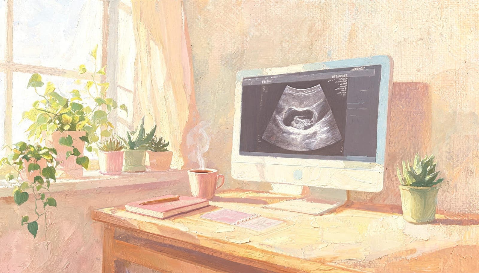
You just had your ultrasound and you're staring at the image, trying to make sense of the gray shapes. Is that a boy? A girl? While definitive gender determination requires a trained professional, understanding the basics of what to look for can be both fun and educational. Here's a parent-friendly guide to reading your ultrasound for gender clues.
Important Disclaimer
Before we begin: reading ultrasounds for gender is not as simple as online forums suggest. What looks obvious to an untrained eye is often misleading. This guide is educational — for a reliable prediction, always consult a professional analyst or your healthcare provider.
Understanding Your Ultrasound Image
The Basics
An ultrasound image uses sound waves to create a visual representation of internal structures. Here's what you need to know:
- Bright/white areas: Dense tissue (bone, cartilage)
- Dark/black areas: Fluid (amniotic fluid, bladder)
- Gray areas: Soft tissue (organs, muscle, skin)
- Top of image: The ultrasound probe/skin surface
- Bottom of image: Deeper structures
Image Orientation
Ultrasound images can be displayed in different orientations, which is one reason DIY gender reading goes wrong:
- Sagittal view: Side profile of the baby (like a silhouette)
- Transverse view: Cross-section (like slicing a loaf of bread)
- Coronal view: Front-facing view (like looking at the baby face-on)
For gender prediction, the sagittal (profile) view is most useful for Nub Theory, and the transverse view is needed for Ramzi Theory.
Method 1: Nub Theory — Reading the Angle
Nub Theory is the most popular early gender prediction method. Here's what to look for:
What Is the Nub?
The "nub" is the genital tubercle — a small protrusion visible between the baby's legs on a sagittal (profile) ultrasound. At 11–14 weeks, all babies have a nub, regardless of gender. The angle of this nub relative to the spine is what predicts gender.
How to Find the Nub on Your Ultrasound
- Find the profile view: Look for an image where you can see the baby's head, spine, and body in profile (like a silhouette)
- Follow the spine down: Trace the baby's spine from head to bottom
- Look at the tailbone area: Just below the base of the spine, you'll see a small bump pointing outward — that's the nub
Reading the Angle
- Boy prediction: The nub angles upward at more than 30 degrees from the line of the spine (pointing toward the head)
- Girl prediction: The nub is parallel to the spine or angles downward (less than 10 degrees)
- Inconclusive: Angle between 10–30 degrees — too early or unclear to determine
Common Mistakes
- Confusing the umbilical cord with the nub (the cord floats freely and changes position)
- Reading at the wrong gestational age (before 11 weeks, the nub hasn't differentiated)
- Wrong image plane (the nub is only visible on a true sagittal view)
Method 2: The Potty Shot — Classic Gender ID
The "potty shot" is the image most people associate with gender determination. It's taken from below, looking up between the baby's legs.
What to Look For
Boy Indicators
- Three visible lines in a dome shape: The scrotum and penis form a characteristic shape that some describe as a "turtle" or "snail"
- Protruding tissue: A visible bump between the legs
- Turtle sign: A round shape (scrotum) with a small extension (penis)
Girl Indicators
- Three parallel lines: The labia appear as three white parallel lines stacked horizontally
- Hamburger sign: The labia and clitoris form a shape that resembles a hamburger bun
- Flat or slightly indented: No protruding tissue between the legs
When the Potty Shot Works
The potty shot is most reliable at 16–20 weeks when external genitalia are fully developed and visible. Before 16 weeks, the structures are too small and similar in appearance for reliable identification.
Method 3: Skull Theory — Reading Facial Structure
Skull Theory suggests that male and female babies have differently shaped skulls. Here's what proponents look for:
Male Skull Features
- Larger, more prominent brow bone (supraorbital ridge)
- Squared jaw
- Steeper forehead
- More angular features overall
Female Skull Features
- Smoother, rounder forehead
- Softer jaw line
- Wider, rounder cranial shape
- Less prominent brow bone
Reality Check
Skull Theory is less scientifically validated than Nub Theory. The differences are subtle, and many experts believe fetal skull shapes are too similar at this stage for reliable differentiation. It's best used as a supplementary method alongside Nub Theory.
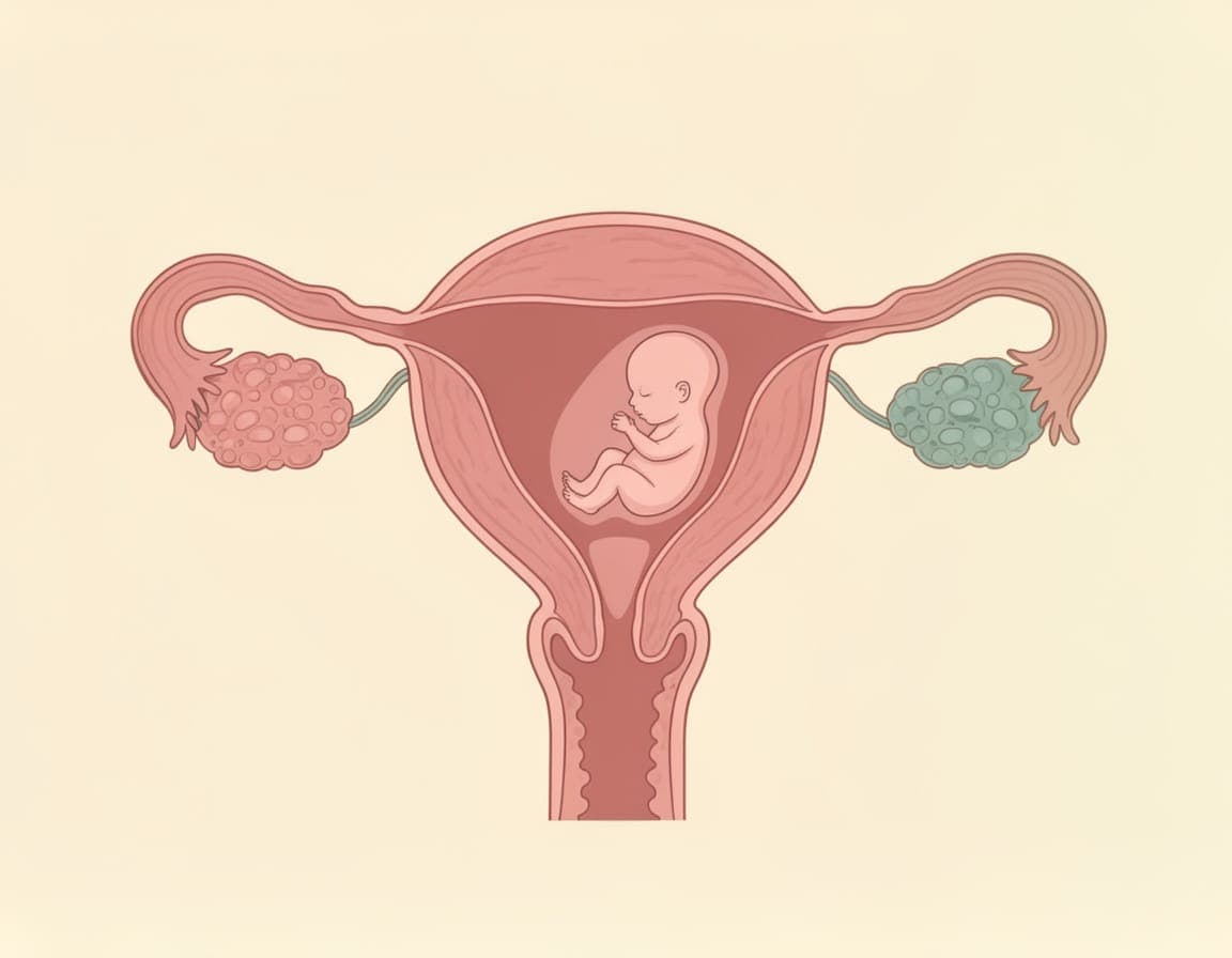
Method 4: Ramzi Theory — Reading Placenta Position
Ramzi Theory is completely different from the above methods — it looks at where the placenta is positioned, not the baby.
How to Read It
- Get a transverse (cross-section) view of your uterus
- Identify the placenta: The bright, thick area attached to the uterine wall
- Determine left vs. right:
- If the placenta is on the right side → predicted boy
- If the placenta is on the left side → predicted girl
Critical Warning About Mirroring
This is where most DIY readings fail:
- Transvaginal ultrasound: NOT mirrored — left on screen = left in body
- Transabdominal ultrasound: MIRRORED — left on screen = right in body
If you don't know which type of ultrasound your image is from, you cannot reliably determine left vs. right.
Why Professional Analysis Beats DIY
Understanding these methods is fun and educational, but there are important reasons to seek professional analysis:
1. Image Interpretation Expertise
Professionals have studied thousands of ultrasounds and can distinguish between:
- The nub vs. the umbilical cord
- The placenta vs. other tissue masses
- Real structural features vs. ultrasound artifacts and shadows
2. Multiple Method Cross-Referencing
Rather than relying on a single method, professional analysts use all available methods and cross-reference results. When multiple methods agree, confidence is much higher.
3. Honest Uncertainty
A good analyst will tell you when your image quality isn't sufficient for a reliable prediction, rather than making a guess that could be wrong.
4. Detailed Explanations
Professional reports explain the reasoning behind the prediction, showing you exactly what was seen and why, with confidence levels for each method.
Getting the Best Ultrasound Images for Gender Prediction
If you're planning to have your ultrasound analyzed:
Ask Your Technician For:
- A sagittal profile view (for Nub Theory)
- A transverse view showing the uterus (for Ramzi Theory)
- A clear between-the-legs view (for potty shot, if past 16 weeks)
- Digital images (higher quality than printed photos)
- Images where the baby is still and clearly visible
Avoid:
- Blurry or dark images
- Images where the baby is in motion
- Photos of photos (re-photographing printed ultrasheets reduces quality)
- Images without any orientation reference
Ready for Professional Analysis?
Now that you understand the basics, let a trained analyst provide a detailed, reliable gender prediction report. Upload your ultrasound and receive results using Nub, Ramzi, and Skull theory — starting at $9.99 with results in 24–48 hours.
Get Your Professional Gender Prediction →
For more helpful resources during your pregnancy, explore our free pregnancy tools — including a due date calculator, ovulation calculator, and gender quiz.
References
- American College of Obstetricians and Gynecologists (ACOG). "Ultrasound Examinations." acog.org, 2024.
- American Institute of Ultrasound in Medicine (AIUM). "Standards for Performance of the Antepartem Obstetric Ultrasound Examination." 2023.
- Efrat, Z., et al. (2006). "Early determination of fetal gender by ultrasound." Ultrasound in Obstetrics & Gynecology, 28(3), 294-297. PubMed
Medical Disclaimer: Gender prediction methods including Ramzi, Nub, and Skull theory are for informational and entertainment purposes only. They are not medically validated diagnostic tools. Always consult your healthcare provider for confirmed medical information about your baby's gender.
Get Your Professional Prediction
Let our experts analyze your ultrasound using the methods discussed in this article.
View Our Services