Gender Prediction at 17 Weeks: Accuracy, Methods, and What to Expect
Gender Prediction at 17 Weeks: Accuracy, Methods, and What to Expect
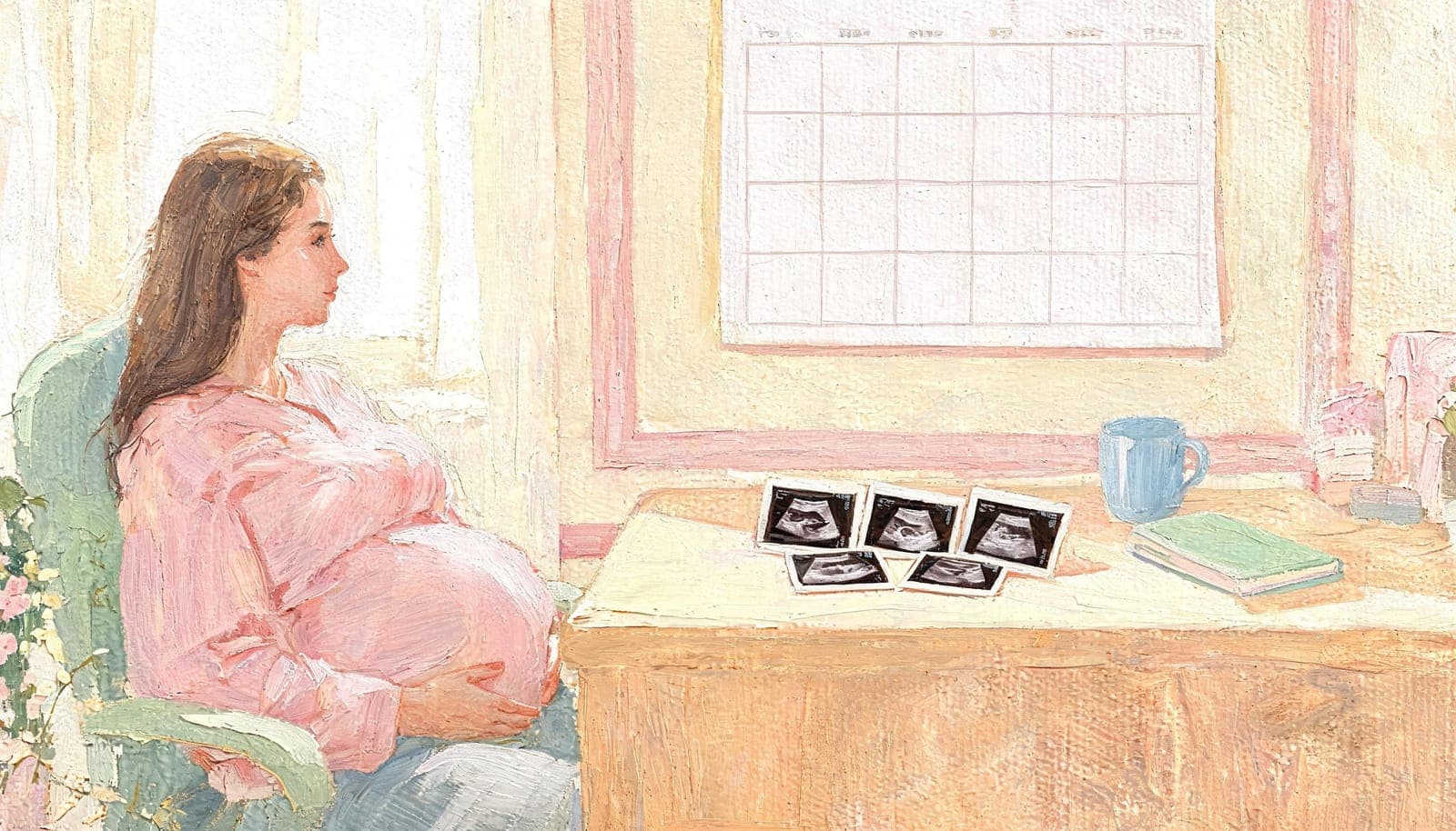
Seventeen weeks pregnant and counting down to finding out whether you're having a boy or a girl? You're in a great position. At 17 weeks, your baby has grown to about 5.1 inches (13 cm) from crown to rump and weighs roughly 5 ounces. The genitalia are well-formed, cranial features are increasingly defined, and ultrasound images at this stage carry more detail than at any earlier point in the second trimester.
Whether you just had your scan or you're about to, here's everything you need to know about gender prediction at 17 weeks.
Not sure of your exact gestational age? Use our free How Far Along Am I? calculator to pinpoint your precise week and trimester.
Why 17 Weeks Is a Strong Window for Gender Prediction
At 17 weeks gestation, several anatomical developments align to make gender prediction especially reliable:
- External genitalia are fully differentiated. By 17 weeks, male and female anatomy is clearly distinguishable on a quality ultrasound. The penis and scrotum in boys and the labia in girls are well-defined structures.
- The nub has completed its transformation. The genital tubercle that nub theory analyzes has fully developed into either male or female anatomy, making this one of the last weeks where nub-based analysis remains highly applicable.
- Cranial bones are more ossified. Skull theory benefits from increasingly pronounced jawlines, brow ridges, and cranial proportions at this stage.
- The placenta is clearly positioned. For Ramzi theory, the placenta's location is unambiguous at 17 weeks, with no confusion between early formation stages.
This combination of factors places 17 weeks in a sweet spot between early theory-based prediction and the definitive 20-week anatomy scan.
Methods Available at 17 Weeks
Nub Theory at 17 Weeks
While the classic nub theory is most discussed at 12-14 weeks, the principles remain relevant at 17 weeks. The genital region continues to display gender-specific characteristics:
- Male indicators: A visible upward angle from the horizontal plane of the lower spine, with a more pronounced protrusion
- Female indicators: A flatter or slightly downward-angled profile with parallel lines
At 17 weeks, however, the "nub" itself has largely completed its development into recognizable genitalia, so analysis may shift from angle measurement to direct feature identification.
Expected accuracy at 17 weeks: 88-95%
This is higher than at 12-14 weeks because the genital structures have fully differentiated. A trained analyst can identify gender characteristics with greater confidence at this stage.
Ramzi Theory at 17 Weeks
Ramzi theory uses placenta position to predict gender:
- Right-side placenta correlates with male pregnancies
- Left-side placenta correlates with female pregnancies
At 17 weeks, the placenta is fully mature and its position is clearly visible on ultrasound, reducing the ambiguity that can affect predictions at earlier gestational ages.
Expected accuracy at 17 weeks: 55-65%
While the placenta is easier to identify at 17 weeks than at 7-10 weeks, Ramzi theory itself has not been consistently validated in clinical literature. It works best as one data point alongside other methods rather than a standalone predictor.
Skull Theory at 17 Weeks
Skull theory analyzes cranial shape and facial bone structure:
- Boy indicators: Squared jaw, pronounced brow ridge, larger cranial vault, thicker skull bones
- Girl indicators: Rounded jawline, smoother forehead, more tapered chin, rounder cranial shape
Expected accuracy at 17 weeks: 70-80%
At 17 weeks, cranial bones have continued to ossify and facial features are more pronounced than at 16 weeks. This gives analysts more structural detail to evaluate, improving reliability compared to earlier weeks.
Direct Visualization
At 17 weeks, direct visualization of the genitalia is increasingly possible, especially with high-quality ultrasound equipment. The potty shot — a view captured from between the baby's legs — can reveal:
- Boy: A visible penis and scrotum (sometimes called the turtle sign)
- Girl: Three parallel lines representing the labia (the hamburger sign)
This direct observation method does not rely on "theories" and, when the image is clear and the baby is in a favorable position, can be very reliable.
Combining Methods for Maximum Accuracy
The strongest gender predictions at 17 weeks come from applying multiple methods simultaneously and cross-referencing results. Our Full Comprehensive analysis applies all applicable techniques:
| Combination | Confidence Level |
|---|---|
| All three theories agree + direct visualization | Very High (92-97%) |
| All three theories agree | High (87-94%) |
| Two theories agree, one ambiguous | Moderate-High (77-87%) |
| Theories conflict | Low-Moderate (60-72%) |
| Only one method provides clear result | Low-Moderate (55-70%) |
At 17 weeks, when methods agree, confidence levels reach their highest point in the pre-anatomy-scan window.
17 Weeks vs. Other Weeks: How It Compares
| Week | Nub Theory | Ramzi Theory | Skull Theory | Direct View | Overall |
|---|---|---|---|---|---|
| 12 weeks | Yes | Yes | Limited | Rarely | Nub + Ramzi |
| 14 weeks | Yes | Yes | Yes | Sometimes | All three |
| 16 weeks | Yes | Yes | Yes | Sometimes | All + direct |
| 17 weeks | Yes | Yes | Yes | Yes | All + direct |
| 20 weeks | Diminishing | Yes | Yes | Yes | Anatomy scan |
Seventeen weeks stands out because all four methods are viable simultaneously. Unlike earlier weeks where some theories produce ambiguous results, 17 weeks offers the most complete toolkit of analysis methods before the standard anatomy scan.
What to Look For in Your 17-Week Ultrasound
If you're preparing for a 17-week scan or reviewing images you already have, here's what matters most for gender prediction:
Best Image Types
- Sagittal views (side profile) are ideal for nub and skull analysis
- Transverse views (cross-section) reveal placenta position for Ramzi theory
- Potty shots (between-the-legs angle) enable direct genital visualization
- Multiple angles give analysts more data points and improve prediction accuracy
Factors That Affect Accuracy
Even at 17 weeks, several variables can influence how reliable a prediction is:
- Baby's position: A breech or posterior-facing baby can hide the genital region from the ultrasound beam
- Image clarity: Grainy, dark, or overexposed images reduce the detail available for analysis
- Amniotic fluid volume: Both too much (polyhydramnios) and too little (oligohydramnios) can affect image quality
- Maternal tissue density: In some cases, body tissue can affect ultrasound penetration
- Equipment quality: Higher-end ultrasound machines capture significantly more detail than basic models
Should You Predict at 17 Weeks or Wait?
Reasons to Predict at 17 Weeks
- You have a clear ultrasound image and want an early answer
- You're planning a gender reveal before your 20-week anatomy scan
- You want to start preparing — nursery themes, names, shopping
- You understand these are theory-based predictions for entertainment purposes
- You want the highest possible accuracy before the anatomy scan window
Reasons to Wait
- You need a definitive medical answer (wait for the anatomy scan)
- Your 17-week image is blurry or poorly angled
- Your healthcare provider has already scheduled an anatomy scan at 18-20 weeks
- You would be significantly upset by an incorrect prediction
Professional Analysis vs. Self-Reading
At 17 weeks, the genital structures are developed enough that you might spot gender clues yourself by comparing your ultrasound to images online. However, professional analysis offers significant advantages:
- Mirroring correction: Abdominal ultrasounds are frequently mirrored (left-right flipped). A trained analyst accounts for this automatically, which is critical for Ramzi theory accuracy.
- Pattern recognition from thousands of scans: What looks ambiguous to untrained eyes may be clearly interpretable by an experienced analyst.
- Honest confidence assessment: A professional will tell you if your image doesn't support a confident prediction rather than guessing to give you an answer.
- Multi-method cross-referencing: Applying all three theories simultaneously and weighing areas of agreement and disagreement produces stronger predictions than any single method alone.
- Detailed written report: You receive a thorough explanation of the analysis methodology and findings, not just a "boy" or "girl" label.
How to Get Your 17-Week Prediction
If you have a 17-week ultrasound image, getting a professional prediction is simple:
- Upload your scan at our secure submission page
- Choose your analysis package — Single Theory ($9.99), Duo Theory Pack ($16.99), or Full Comprehensive ($24.99)
- Provide scan details — Gestational age, scan type (abdominal or transvaginal), and any notes about the image
- Receive your report — A detailed analysis with confidence levels delivered to your email within 24-48 hours
For 17 weeks, we recommend the Full Comprehensive package. Since all three theories are highly applicable at this stage and direct visualization is often possible, combining every available method produces the strongest prediction possible before your anatomy scan.
Upload Your Ultrasound → and get your professional gender prediction report. Starting at $9.99 with results in 24-48 hours.
Medical Disclaimer: Gender prediction using ultrasound theory methods is for informational and entertainment purposes only. These methods are not medically diagnostic. The anatomy scan at 18-20 weeks performed by your healthcare provider is the standard for medical gender determination. Always consult your healthcare provider for confirmed medical information about your baby.
Get Your Professional Prediction
Let our experts analyze your ultrasound using the methods discussed in this article.
View Our Services